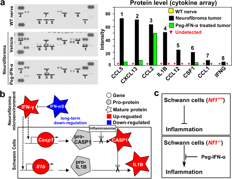
Figure 8. Pro-inflammatory cytokines in Neurofibroma.
(a) Left panel: Pro-inflammatory cytokines are at low levels in wild-type nerve (top), show increased protein levels in neurofibroma (middle), and are reduced after treatment of neurofibroma with PEGylated IFN-α2b (bottom). Right panel: Relative intensity, reflecting comparative levels of expression for each protein, after the intensity of pixels was averaged and plotted. (b) A model developed from gene expression analysis (drawn by Inkscape v0.48, http://inkscape.org). Decreased levels of type-I interferons and increased type-II interferon increase inflammation in the tumor microenvironment by increasing expression of Casp1 and Il1b mRNAs. CASP1 pro-protein is known to be cleaved and thus be activated by the inflammasome. Active CASP1 cleaves pro-IL1B protein, releasing active IL1B cytokine. (c) Based on this analysis, normal SCs suppress nerve inflammation. When Nf1−/− SCs are present, de-regulated interferons result in inflammation, which can be largely normalized by PEGylated IFN-α2b.

