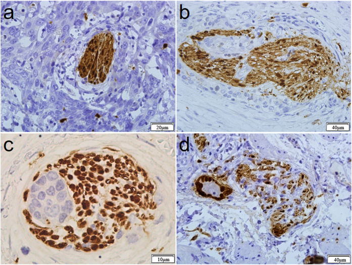
Figure 4. Variety patterns of the histologic appearance of PNI in ESCC.
(a) The nerve fiber was tightly surrounded by tumor cells. The perineurium of the nerve exhibited high integrity. (b) Tumor cells invaded the nerve and damaged the perineurium of the nerve. (c) Tumor cells invaded the nerve. (d) The nerve fibers were irregularly damaged by tumor cells, which broke through the perineurium and grew outward.

