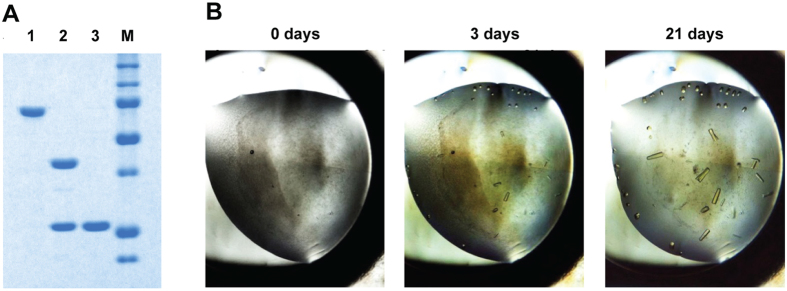
Figure 7. Preparation of ΔABC for crystallisation.
(A) Removal of the MBP-tag and purification of native ΔABC, visualised on SDS-PAGE. From left to right: (1) purified MBP-TtProDH ΔABC; (2) MBP-TtProDH ΔABC after limited trypsinolysis; (3) purified native ΔABC. Molecular masses of marker proteins (M) from top to bottom: 150, 100, 75, 50, 37, 25, 20 kDa. (B) Growth of TtProDH ΔABC crystals.

