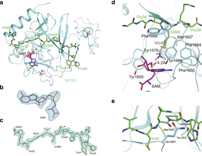
Figure 1. The structure of the SET domain of SETD2 in complex with an H3K36M peptide and SAM.
(a) The overall structure of the SET domain in complex with the H3K36M peptide. The SET domain is shown in cyan, the H3K36M peptide in green stick models and SAM in magenta. Several zinc ions bound to the protein are shown as pink spheres. (b) Omit Fo−Fc electron density at 2.42 Å resolution for SAM, contoured at 3σ. (c) Omit Fo−Fc electron density at 2.42 Å resolution for the H3K36M peptide, contoured at 3σ. (d) Detailed interactions between the SETD2 SET domain (cyan) and the H3K36M peptide (green) near K36M. The distance between the methyl group of SAM and the sulfur atom of K36M is indicated by the dashed line. The bound position of dimethylated H3K9 (labeled Mly9) peptide to GLP125 is also shown (gray). (e) Pro38 assumes a trans configuration and has favorable interactions with the SET domain. All structure figures were produced with PyMOL (www.pymol.org).

