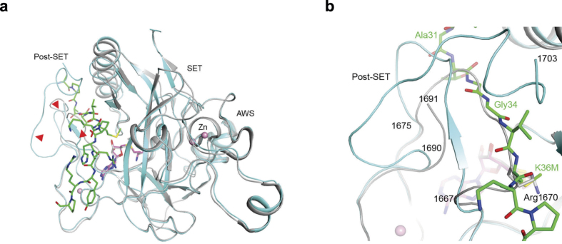
Figure 2. Extensive conformational changes in SETD2 SET domain upon H3K36M peptide and SAM binding.
(a) Overlay of the structure of SETD2 SET domain (cyan) in complex with H3K36M peptide (green) and SAM (magenta) with that of the SET domain in complex with SAH (gray)26. Red arrowheads point to regions of large conformational differences between the two structures, in the post-SET motif. (b) The binding site for the substrate peptide (green) is occupied by a loop of the protein in the free SET domain structure (gray)26, due to large conformational changes for residues 1667–1675 and those at the C-terminus of the SET domain.

