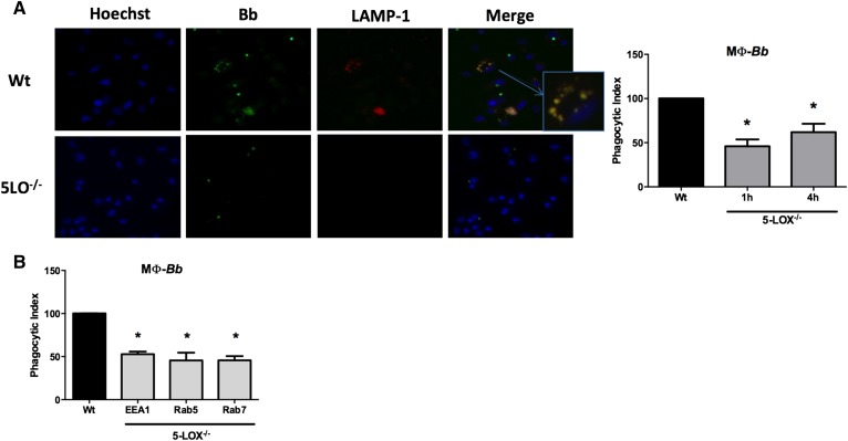
Fig. 1.
Defective phagocytosis in macrophages from 5-LOX−/− mice occurs at early stages of phagosome development. BMDMs from WT or 5-LOX−/− mice were cocultured with GFP-B. burgdorferi (Bb) for 1 or 4 h. A: Images from fluorescence microscopy of WT or 5-LOX−/− macrophages cocultured with GFP-Bb for 4 h and stained with Hoechst (nuclear stain) or LAMP-1 (lysosomal stain). The inset shows colocalization of GFP-Bb and LAMP-1 in macrophage endosomes. The phagocytic index shows the level of uptake of Bb by 5-LOX−/− macrophages (MΦ) as compared with WT macrophages. Because the phagocytosis levels of WT macrophages at 1 and 4 h was arbitrarily set to 100%, only a single WT bar is shown. B: Phagocytic index of 5-LOX−/− macrophages using different markers of phagosome progression (EEA, early endosome; Rab5, early endosome; Rab7, late endosome). Bars represent mean + SEM and are the results from two independent experiments (n = 3 per group). *P < 0.01 versus corresponding WT control cells.

