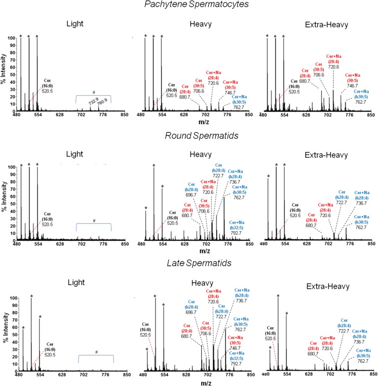
Fig. 7.
MALDI-TOF MS analysis of the Cer species from membrane fractions of differentiating spermatogenic cells. The Cers of the indicated membrane fractions from the spermatogenic cells under study were isolated by TLC and purified as described in the text. All molecular species of Cer were identified by the product ion scan m/z +264 that corresponds to twice-dehydrated sphingosine (d18:1). The n-V Cer and h-V Cer species are colored in red and blue, respectively. Signals at m/z 680.7 and 706.6 are [(M+H)+ − H2O] of (d18:1/28:4n-6)Cer and (d18:1/30:5n-6)Cer, respectively. The latter two species were also detected as sodium adducts (Cer + Na+) at m/z 720.6 and 746.7, respectively. The intensities detected at m/z 696.7 and 722.7 are dehydrated species [(M+H)+ − H2O] of (d18:1/h28:4n-6)Cer and (d18:1/h30:5n-6)Cer, respectively. The latter two molecular species of Cer were also detected as sodium adducts (Cer + Na+) at m/z 736.7 and 762.7, respectively. In light membranes from the three cell types, no Cers with VLCPUFA were present and only minor amounts of 16:0 Cer (m/z 520.5) were detected. The peaks marked with an asterisk indicate matrix ions signals. The area under the # symbol corresponds to unidentified signals. The heavy membranes from spermatids showed a signal at m/z 792.7 that corresponds to (d18:1/h32:5n-6 + Na+)Cer.

