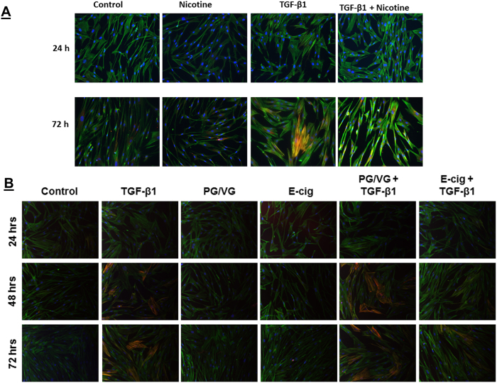
Figure 3. TGF-β1 induced fibroblast to myofibroblast differentiation is blocked by nicotine and e-cig condensate.
Representative immunofluorescence microscopy images of HFL-1 for α-SMA (A) HFL-1 cells treated with TGF-β1, nicotine, TGF-β1 co-treated with nicotine, or no treatment control for 24 hrs or 72 hrs. The cell nuclei were stained with Dapi (blue) and actin with Phalloidin (green). Representative immunostaining of α-SMA-positive filamentous structures (yellow). (B) Treatment for 24 hrs, 48 hrs, or 72 hrs with TGF-β1, PG/VG, E-cig, TGF-β1 co-treated with PG/VG, or TGF-β1 with E-cig. Original magnification, 200x. Data are a representative of at least three reproducible experiments. E-cig, e-cigarette; PG, propylene glycol; VG, vegetable glycerin.

