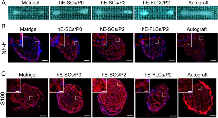
Figure 5. Macroscopic view and immunofluorescence staining of frozen sections of regenerated sciatic nerve tissue 12-weeks after surgery in the five groups.
(A) Macroscopic view (B) anti-NF-H staining, and (C) anti-S100 staining. The white solid-line boxed areas were enlarged in the upper left inset. Images are representatives of the following groups: Matrigel, hE-SCs/P0. hE-SCs/P2 hE-FLCs/P2,and Autograft. Scale bars = 100 μm, scale bars in the upper left inset = 25 μm.

