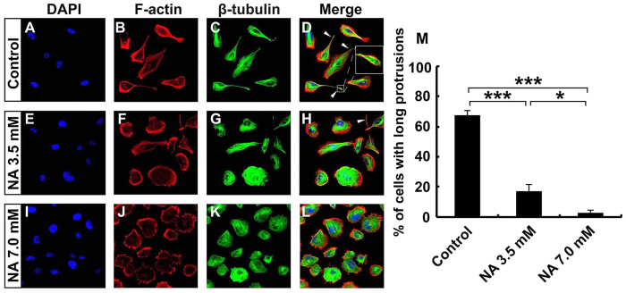
Figure 1. U251 cells lose mesenchymal phenotype upon NA treatment.
U251 cells were incubated with PBS (control) or the indicated concentration of NA for 8 hr. Rhodamine phalloidin labeling for F-actin (red), immunocytochemistry for β-tubulin (green) and DAPI labeling for nuclei (blue) were carried out as described in Methods. White arrowheads denote the long protrusions that consist mainly of microtubules, and the inset in (D) shows an amplified image of the indicated protrusion. The percentage of cells with this type of long protrusions was calculated for each treatment and summarized in (F). *P < 0.05; ***P < 0.001.

