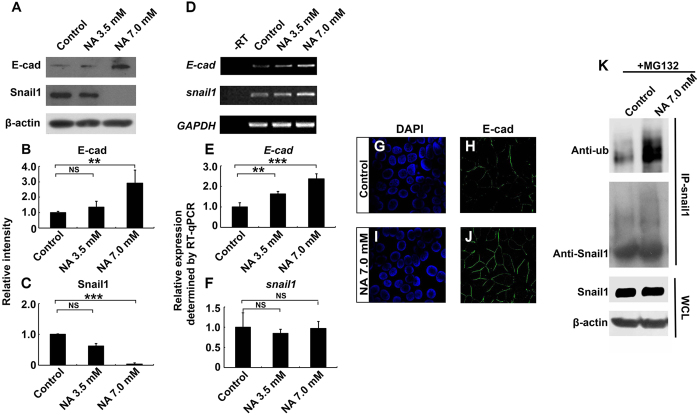
Figure 3. NA upregulates E-cadherin expression by promoting ubiquitination and degradation of Snail1.
U251 cells were treated with PBS (control) or the indicated concentration of NA for 4 hr. (A–C) Western blot analyses for whole-cell lysates were performed with an anti-E-cadherin antibody. Membranes were stripped and reblotted for Snail1 and β-actin. Representative images of Western blots are shown in A, and relative intensity of E-cadherin and Snail1 normalized against β-actin was calculated and summarized in (B,C), respectively. (D–F) Total RNA was extracted, and RT-PCR was carried out for the transcripts of E-cadherin, snail1, and GAPDH. Representative images of semi-quantitative RT-PCR are shown in (D) and relative expression levels of E-cadherin and snail1 (normalized against GAPDH), as determined by RT-qPCR, are shown in (E,F) respectively. NS, not significant; **P < 0.01; ***P < 0.001. (G–J) Cells were fixed and processed for DAPI staining (blue) and immunocytochemistry for E-cadherin (green). (K) U251 cells were treated with MG132 and NA or PBS (control), IP was carried out for cell lysates with an anti-Snail1 antibody, and Western blot was performed with an anti-ubiquitin antibody. Western blot for whole-cell lysates (WCL) was also performed separately with an anti-Snail1 antibody.

