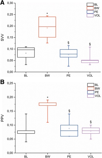
Fig. 2.

Box plots showing changes in A: stroke volume variation (SVV) and B: pulse pressure variation (PPV) during baseline (BL), blood withdrawal (BW), pulmonary embolism (PE) and volume loading (VOL). The line in each box indicates the median. The upper and lower limits of each box indicate the 75th and 25th percentiles, respectively. The error bars above and below each box represent the 90th and 10th percentiles, respectively. * P < 0.05 vs BL; § P < 0.05 vs BW
