Fig. 2.

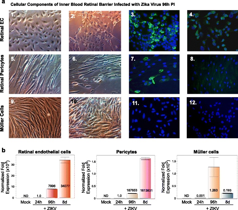
Cellular components of the inner blood-retinal barrier and ZIKV infectivity. Phase contrast images of a an uninfected confluent monolayer of retinal endothelial cells (a-1), a confluent monolayer of retinal endothelial cells infected with ZIKV 96 h after infection (a-2), immunofluorescence staining of ZIKV-infected endothelial cells with the Flavivirus 4G2 antibody (a-3), an uninfected confluent monolayer of retinal pericytes (a-4), a confluent monolayer of retinal pericytes infected with ZIKV 96 h after infection (a-5), immunofluorescence staining of ZIKV-infected pericytes with the Flavivirus 4G2 antibody (a-6), an uninfected confluent monolayer of Müller cells (a-7), a confluent monolayer of Müller cells infected with ZIKV 96 h after infection (a-8), and an immunofluorescence staining of ZIKV-infected Müller cells with the Flavivirus 4G2 antibody (a-9). Mock-infected controls of retinal endothelial cells (a-4), retinal pericytes (a-8), and Müller cells (a-12) stained with the 4G2 antibody. All images were taken on a Nikon TE2000S microscope mounted with a charge-coupled device (CCD) camera at ×200 total magnification. For fluorescent images, 4′,6-diamidino-2-phenylindole (DAPI) was used to stain the nuclei blue. b qRT-PCR time course of retinal endothelial cells, retinal pericytes, and Müller cells infected with ZIKV for 24 and 96 h and 8 days after infection. Mock-infected controls are also shown. All values were normalized to GAPDH. ND indicates no transcriptional expression detected
