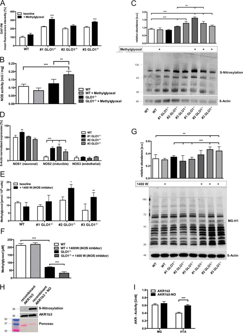
FIGURE 4.
S-Nitrosylation of AKR1b3 is beneficial for the efficient detoxification of dicarbonyl species in GLO1−/− Schwann cells. A, intracellular levels of nitric oxide species in wild-type (□) Schwann cells and three individual GLO1−/− (■) Schwann cell clones using flow cytometry and DAF-FM as a dye reagent. B, enzymatic activity of nitric-oxide synthases in wild-type and GLO1−/− Schwann cells (clone 1), where 1 unit of NOS activity is the amount of enzyme required to yield 1 μmol of nitric oxide/min. C, densitometry analysis and representative Western blot of whole cell lysates of wild-type Schwann cells and three individual GLO1−/− Schwann cell clones with and without MG treatment (50 μm; 6 h) detecting nitrosylated cysteine residues using the iodoTMT switch technique. Lysates were probed with anti-iodoTMT and β-actin as a loading control. D, baseline mRNA expression of the three different subtypes of nitric-oxide synthases (NOS) in wild-type (□) Schwann cells and three individual GLO1−/− (■) Schwann cell clones. Values for NOS1 in wild-type Schwann cells were standardized to 100%. E, intracellular MG levels in wild-type Schwann cells and three individual GLO1−/− Schwann cell clones treated with iNOS inhibitor 1400 W dihydrochloride (100 μm; 24 h). F, median lethal doses (LD50) for MG (48 h exposure time) in wild-type and GLO1−/− Schwann cells (clone 1) treated with iNOS inhibitor 1400 W dihydrochloride (100 μm). G, densitometry analysis and representative Western blot of total cell extracts (30 μg of protein) from wild-type Schwann cells and three individual GLO1−/− Schwann cell clones with and without 1400 W treatment (100 μm; 24 h), probed with anti-MG-H1 antibody detecting MG-modified arginine residues and anti-β-actin antibody as a loading control. H, densitometry analysis and representative Western blot of recombinant AKR1b3 protein with and without exposure to a nitric oxide donor (streptozotocin). Blots were probed with anti-AKR1b3 antibody and stained with Ponceau S staining as a loading control. I, enzymatic activity assay of recombinant AKR1b3 under substrate saturating conditions (2 mm of each) with (■) and without (□) the exposure to a NO donor, where 1 unit is the amount of AKR1b3 that catalyzes the formation of 1 μmol of NADP in 1 min. All data represent the mean of at least four independent experiments ± S.E. Western blot bar graphs represent the mean of three independent experiments ± S.E. ***, p < 0.0001; **, p < 0.001; *, p < 0.05.

