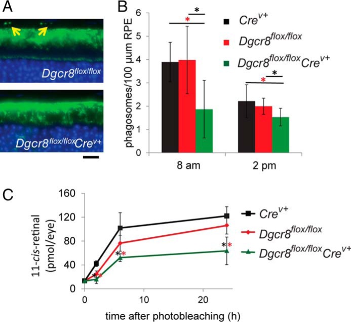
FIGURE 3.

Inducible mature RPE-specific Dgcr8flox/floxCrev+ mice are defective in phagocytosis of photoreceptor outer segment discs and regeneration of visual chromophore. A, immunostaining with an anti-opsin antibody shows foci in the RPE layer (indicated by yellow arrows) that correspond to phagocytosed rod outer segment discs in 8-week-old mice. Scale bar, 25 μm. B, quantitation of the number of phagosomes in Dgcr8flox/floxCrev+ animals near the morning peak and afternoon trough of circadian regulated RPE phagocytosis revealed a reduction in the quantity of opsin-containing foci in the RPE of 8-week-old Dgcr8flox/floxCrev+ mice compared with control animals. p values were <0.001 calculated for Dgcr8flox/floxCrev+ versus Dgcr8flox/flox (black asterisks) and Dgcr8flox/floxCrev+ versus Crev+ (red asterisks). C, analysis of the rate of recovery of the visual chromophore 11-cis-retinal after illumination in 5-week-old Dgcr8flox/floxCrev+ mice and control animals revealed reduced chromophore levels in cKO animals at all studied time points. Error bars, S.D. based on quantification of at least 5 animals/genotype at each time point.
