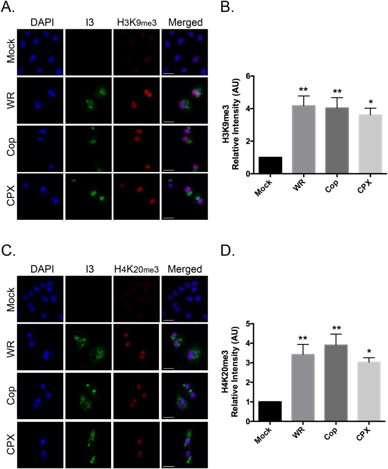
Fig 3. Orthopoxviruses promote H3K9me3 and H4K20me3 formation.
BSC-40 cells were grown on coverslips and subsequently infected with VACV WR, VACV Cop, and CPXV. The cells were fixed and stained to detect I3 and (A) H3K9me3 or (C) H4K20me3 9hr post-infection. DNA was counterstained with DAPI. Images were acquired at 60x magnification (scale bar = 25 μm). The nuclear (B) H3K9me3 and (D) H4K20me3 signal intensities were quantified using FIJI imaging analysis software and normalized to mock-infected cells. Data represent the SEM of three independent experiments and any statistically significant differences relative to mock-infected cells, are noted (*P<0.05; **P<0.01).

