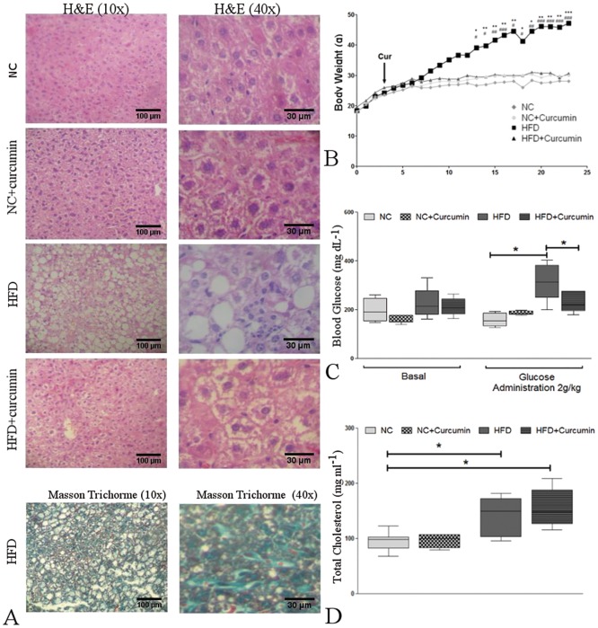
Fig 6. HFD-induced metabolic and histological alterations in the liver and the reversal effects of curcumin.
(A) Top: Hematoxylin and eosin staining of liver sections from normal chow (NC)-fed and high-fat diet (HFD)-fed mice with or without curcumin supplementation (10X and 40X magnification). Curcumin prevented HFD-induced steatosis, ballooning and liver inflammation. Bottom: Masson's trichrome staining of liver sections from HFD-fed mice reveals signs of mild perisinusoidal fibrosis (10X and 40X magnification). (B) Body weight gain in NC- and HFD-fed mice with or without curcumin supplementation, starting at week 4 (arrow) (*p<0.05, **p<0.01 and ***p<0.001, HFD vs. NC). Curcumin prevented HFD-induced weight gain (#p<0.05, ##p<0.01 and ###p<0.001, HFD vs. HFD+curcumin). (C) The blood glucose concentrations were measured upon conclusion of the dietary treatments, and glycemia was determined at basal conditions (Basal) and after glucose administration. Left: a similar blood glucose concentration was observed among the four groups. Right: hyperglycemia was observed in HFD-fed mice at 120 min after an intraperitoneal glucose injection (2 g/kg). Curcumin ameliorated the hyperglycemic conditions. (*p<0.01). (D) Total serum cholesterol levels were measured upon conclusion of the dietary treatments. The HFD induced higher levels of cholesterol than the normal chow regardless of curcumin administration (*p<0.05). The box and whiskers show the non-parametric statistics: the median, lower and upper quartiles and confidence interval around the median. The Kruskal-Wallis test with Dunn’s post-test was performed.

