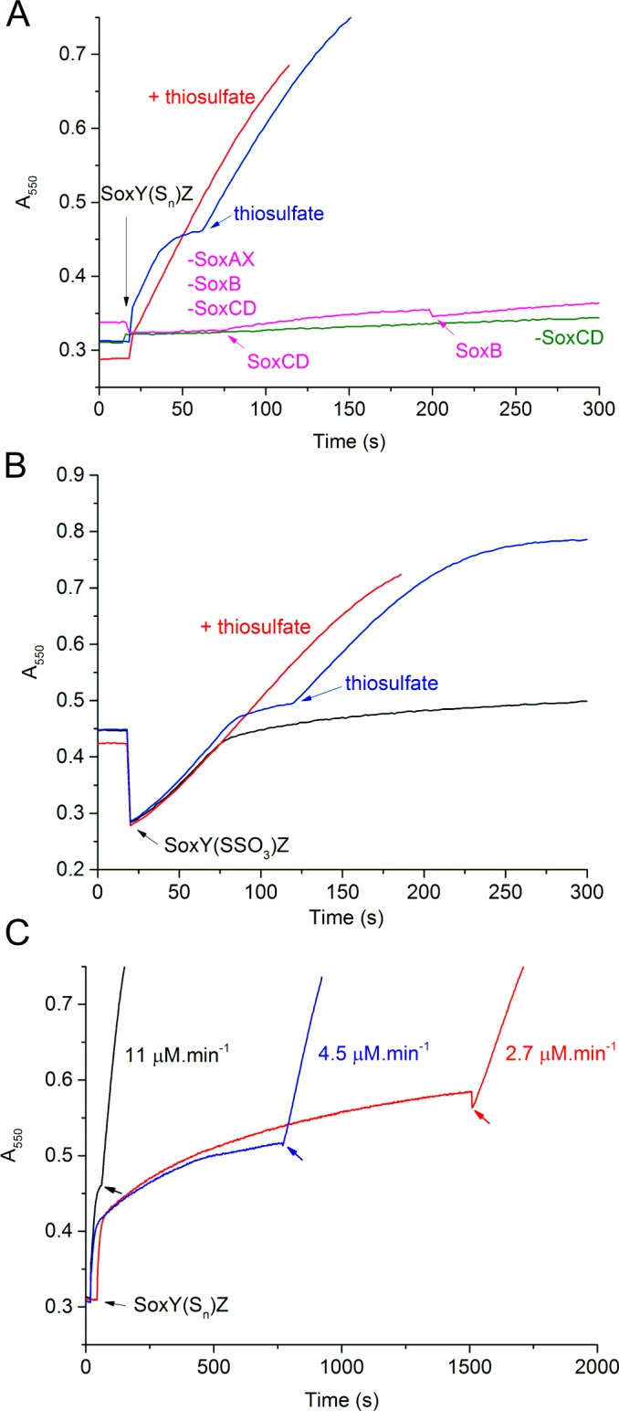
Fig 4. Thiosulfate-independent cytochrome c reduction.

Reactions contained 0.1μM SoxB, SoxCD and SoxAX, unless indicated, and 35 μM cytochrome c. Any missing Sox components are indicated by a minus sign (e.g. `-SoxCD’). Reactions were initiated in panels (A) and (C) by the addition of 1 μM tetrathionate-then-sulfide-treated SoxYZ from fraction 3 of the IEX separation shown in Fig 3C (designated `SoxY(Sn)Z’), or in panel (B) by the addition of 10 μM tetrathionate-treated SoxYZ from fraction 3 of the IEX separation shown in Fig 3B (designated `SoxY(SSO3)Z’). Where indicated, Sox components or thiosulfate were added to final concentrations of 0.1 μM or 2 mM, respectively. The progress of the reactions was assessed through monitoring the reduction of cytochrome c at 550nm.
