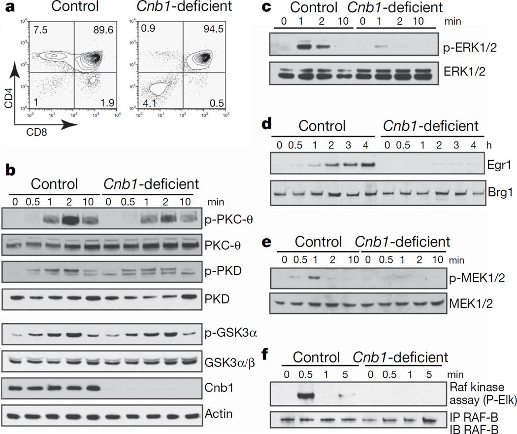
Figure 1. Specific and severe defect in Raf–MEK–ERK activation in Cnb1-deficient thymocytes.
a, Expression of CD4 and CD8 on Cnb1-deficient and control thymocytes. The numbers in the corners of the panels represent the percentage of cells in each quadrant. b, Immunoblot analysis of phosphorylated and total proteins in Cnb1-deficient and control double-positive thymocytes after CD3ε crosslinking. GSK, glycogen synthase kinase; PKC, protein kinase C; PKD, protein kinase D. c, Immunoblot analysis of phosphorylated ERK1/2 in Cnb1-deficient and control double-positive thymocytes after CD3ε crosslinking. d, Immunoblot analysis of Egr1 induction in double-positive thymocytes from Cnb1-deficient and control littermates. Brg1 shows equal loading. e, Immunoblot analysis of phosphorylated MEK1/2 in Cnb1-deficient and control double-positive thymocytes after CD3ε crosslinking. f, Raf-B kinase activity in Cnb1-deficient and control double-positive thymocytes after CD3ε crosslinking. IP, immunoprecipitation; IB, immunoblotting.

