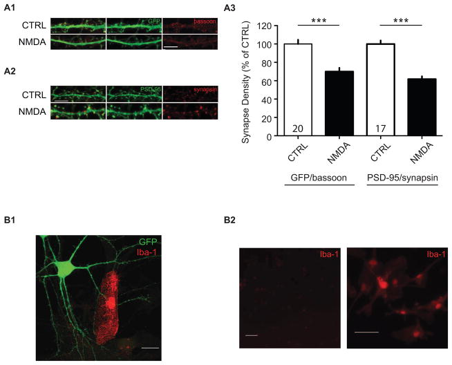
Figure 3. NMDA-induced synapse loss is not likely to be due to loss of synapsin or GFP.
NMDA-induced synapse loss is still observed using an alternative presynaptic marker, bassoon (A1), or postsynaptic marker, PSD-95 (A2). Confocal images after 0 (CTRL) and 2 NMDA treatments showing fluorescent immunoreactive staining using alternative markers of (A1) the presynaptic bouton (red, bassoon), and (A2) the postsynaptic spine, PSD-95. Scale bar, 5 μm. (A3) Quantification of normalized synapse density from neurons stained with alternative antibody combinations, anti-GFP/anti-bassoon, and anti-PSD95/anti-synapsin. Data are averaged means of synapses from at least three independent experiments. Values within bars represent sample sizes (# neurons). Error bars represent SEM. Significance from control (CTRL): *** p < 0.001. (B1) Synapse pruning is unlikely to be a result of microglia action. Example image of an Iba1-positive microglial cell contacting a GFP-expressing neuron. Scale bar, 20 μm. (B2) Relative Iba-1-immunoreactivity in neuronal cultures using our neuron culture protocol (left), and microglia-enriched cultures grown in serum-containing media (Iba1-positive control, right). Scale bars, 50 μm.

