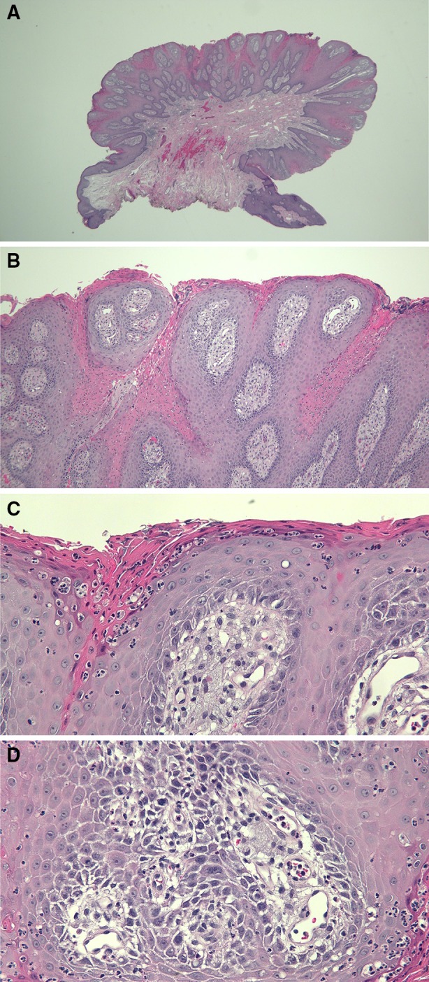
Fig. 2.

Microscopic examination of the lesion from the 83-year-old man was performed. Low magnification (a) shows a pedunculated tumor with acanthosis, papillomatosis, and elongation of the rete ridges. Intermediate magnification (b, c) reveals parakeratosis and neutrophilic inflammation in the dermis. High magnification (d) reveals numerous foamy histiocytes in the widened dermal papillae. Correlation of the clinical features and the pathologic changes establish a diagnosis of verruciform xanthoma. The lesion was completely removed at the time of biopsy, and the patient applied mupirocin 2% ointment to the site. The excision site has since completely healed without recurrence. (Hematoxylin and Eosin: a = ×2, b = ×10, c = ×20, d = ×40)
