
Figure 7.
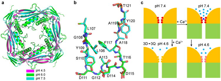
Structures of the TRPML1 I-II linker at different pH and model of Ca2+/pH regulation. (a) Superposition of the I-II linker crystal structure obtained at pH 4.5, 6.0 and 7.5, viewed from the extracellular/luminal side of the membrane. (b) Side view of the superimposed luminal pore-loop structures obtained at pH 4.5, 6.0, and 7.5. (c) Model of Ca2+/pH dual regulation of TRPML1. Two of the four channels are schematized, with yellow representing the ED and green the TMD. The luminal pore aspartates are illustrated in red or pink, depending on luminal pH, and in yellow when mutated to glutamine. Light blue dots represent free Ca2+ ions, and the dark blue dot represents a bound Ca2+ ion. See text for details.