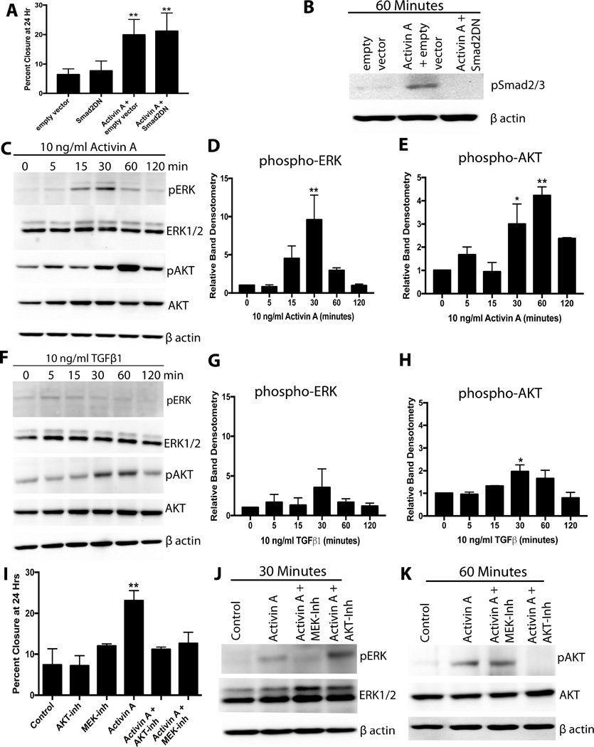
Figure 2.
PI3K/AKT and MEK/ERK but not Smad2/3 pathways are required for activin A-stimulated migration of MOE cells. A) Migration of MOE cells transfected with a dominant negative Smad2 (Smad2DN) construct, empty vector, and treated with 10 ng/ml activin A as indicated. B) Western blot for phospho-Smad2/3 in MOE cells treated activin A and Smad2DN construct as indicated. C–E) Example western blots (C) and densitometry for phospho-ERK (D) and phospho-AKT (E) of MOE cells treated with 10 ng/ml activin A. F–H) Example western blot (F) and densitometry of phospho-ERK (G) and phospho-AKT (H) of MOE cells treated with 10 ng/ml TGFβ1. I) Migration of MOE cells treated with activin A (10 ng/ml), an AKT inhibitor (MK2206), or a MEK inhibitor (U0126) as indicated. J and K) Example western blots for phospho-AKT and phospho-ERK in MOE cells treated with activin A for 30 (J) or 60 (K) minutes in the presence of an AKT inhibitor (MK2206) or an ERK inhibitor (U0126). *P<0.05, **P<0.01, n≤3.

