Figure 6.
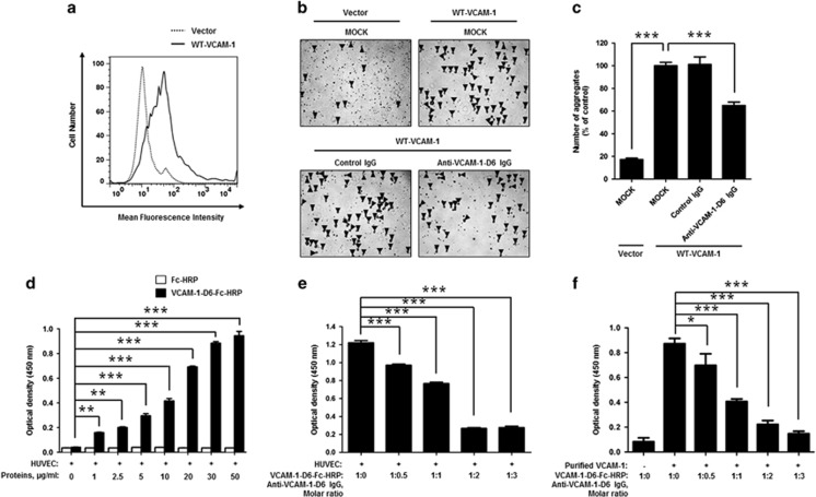
Effect of anti-VCAM-1-D6 IgG on clec14a-mediated cell–cell contact in VCAM-1-overexpressing HEK293F cells and hTNFα-treated HUVECs. (a) HEK293F cells were transfected with vector alone or expression plasmid encoding WT-VCAM-1. Cells were stained with anti-VCAM-1 monoclonal antibody and VCAM-1 expression was analyzed using flow cytometry. (b) HEK293F cells transfected with vector alone or expression plasmid encoding WT-VCAM-1 were incubated in the absence (MOCK) or presence of 20 μg ml−1 control IgG or anti-VCAM-1-D6 IgG. Cell aggregates (masses >4 cells; triangular arrowheads) were counted under a light microscope. (c) Quantitation of aggregates formed in the transfected cells in the absence (MOCK) or presence of control IgG or anti-VCAM-1-D6 IgG expressed as a percent of control (MOCK) aggregate formation. (d) ELISA results of binding of the indicated concentrations of anti-VCAM-1-D6-Fc-HRP and Fc-HRP to hTNFα-treated HUVECs. (e) ELISA results of VCAM-1-D6-Fc-HRP binding, in the absence or presence of the increasing concentrations of anti-VCAM-1-D6 IgG, to hTNFα-treated HUVECs pre-incubated with 3 μg ml−1 VCAM-1-D6-Fc-HRP. (f) One tenth of a microgram of VCAM-1-D6-Fc-HRP was pre-incubated with 1 μg recombinant human VCAM-1 extracellular domain, and binding was determined by ELISA in the absence or presence of the increasing concentrations of anti-VCAM-1-D6 IgG. All data are presented as the mean±s.e.m. of triplicate measurements from one of three independent experiments; *P<0.05, **P<0.01, ***P<0.001.

