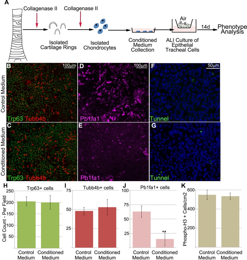
Fig. 2.
Chondrocyte-conditioned medium inhibits club cell differentiation. A: concept diagram of the protocol for chondrocyte isolation and differentiation of tracheal epithelial cells. ALI, air-liquid interface. B–E: analysis of respective population differences between control and conditioned cells by fluorescence imaging of ALI cell cultures. Cells fed conditioned medium from chondrocytes showed no significant change in frequency between basal [transformation-related protein 63-positive (Trp63+)] and ciliated [tubulin-β4B, class IVb (Tubb4b+)] cells and control cells. However, the number of club (Pb1fa1+) cells was significantly decreased in chondrocyte-conditioned medium. F and G: no increase in apoptotic rate in tracheal cells cultured with chondrocyte-conditioned medium. Images are representative of 4 different samples. H–J: quantification of results from fluorescence imaging (B–E) illustrating reduced number of club (Pb1fa1+) cells, but no change in the number of basal or ciliated cells, fed chondrocyte-conditioned medium. K: no statistically significant difference in proliferation or apoptosis between cells fed chondrocyte-conditioned medium and cells fed control medium. phospho-H3, phosphorylated histone. **P < 0.01. Values are means ± SD of 4 separate experiments.

