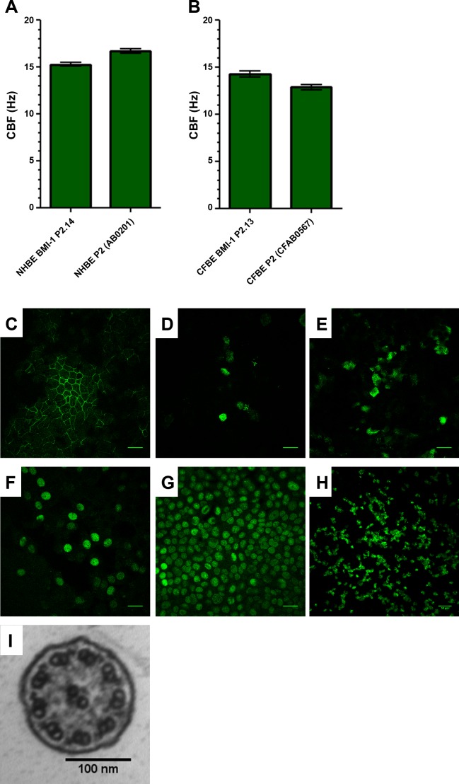
Fig. 4.
BMI-1 cells retain their mucociliary differentiation capacity. Extensively passaged BMI-1-transduced cells (passage 15) were differentiated on ALI and cilia beat frequency of NHBE (A) and CFBE (B) cells was determined using ciliaFA plugin (25) for ImageJ. Data are means ± SE; n = 4 independent ALI cultures, 5 fields videoed per culture. Immunostaining of NHBE-BMI-1 cells was used to show tight junction formation (occludin; C), mucin production (MUC5AC and MUC5B; D and E, respectively), the presence of basal cells (p63+; F), widespread BMI-1 expression (BMI-1; G), and extensive ciliation (acetylated α-tubulin; H). TEM was used to determine cilia ultrastructure (I). Images are representative of 4 independent ALI cultures per marker. Scale bars for C–H are 50 µm and 100 nm for I.

