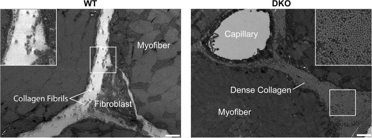
Fig. 1.
Extensive fibrosis is present in double knockout (DKO) skeletal muscle. Transverse transmission electron micrographs of the extracellular space of wild-type (WT) and nesprin-1/desmin DKO (13) tibialis anterior muscle show a significant increase in collagen content in DKO muscle. Inset shows that DKO collagen is more densely packed compared with WT collagen, with collagen fibrils roughly aligned with the muscle fiber long axis. Scale, 500 nm.

