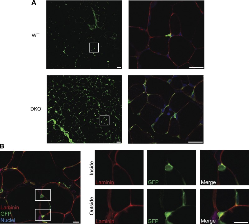
Fig. 2.
Morphological evidence of increased type I collagen-expressing cells. A: collagen-expressing cells are more abundant in DKO skeletal muscle sections compared with WT. White boxes in the left panel correspond to the area in the right panel. In addition to an increased number of collagen expressing cells in DKO muscle, high-magnification images reveal an increase in cellular projections of collagen-expressing cells in DKO muscle. Scale, 25 μm. B: collagen-expressing cells (GFP+) were found both inside and outside of the muscle cell. White boxes in left figure depict enlarged areas on the right. In right figure, the top panel clearly shows a GFP+ collagen-expressing cell lying completely beneath the basal lamina. The bottom panel illustrates a collagen-expressing cell outside of the basal lamina nestled between two muscle fibers. This shows that collagen-expressing cells are a heterogeneous cell population. Scale, 10 μm. Red = laminin, green = GFP+ collagen cells and blue = DRAQ5 nuclear staining.

