Fig. 4.
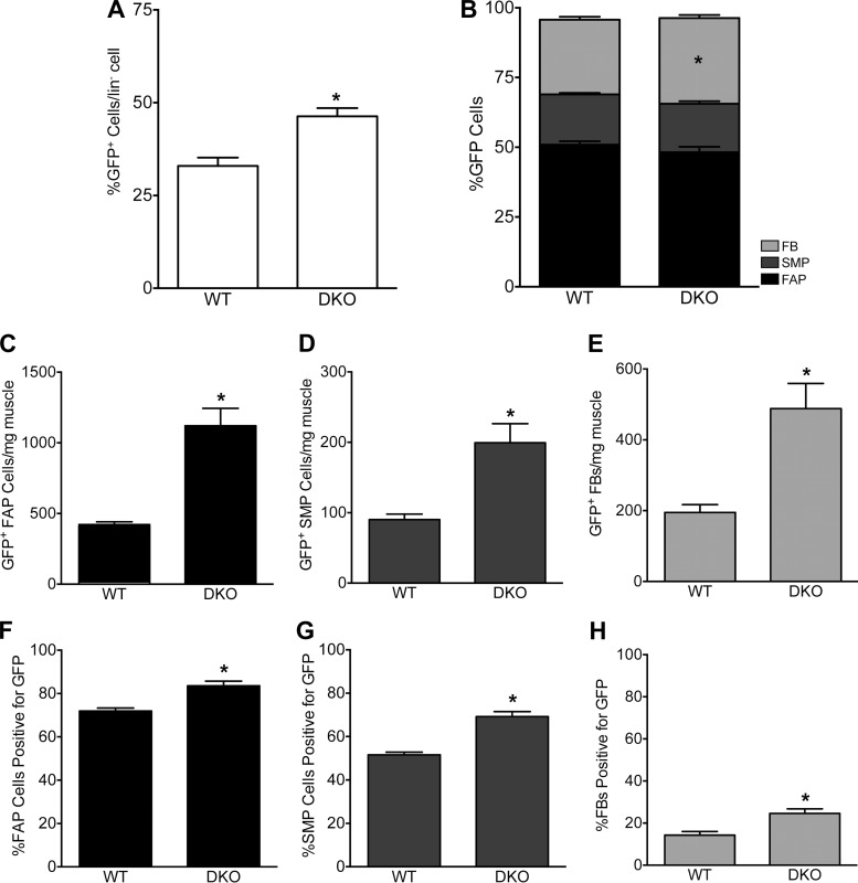
Multiple collagen I-expressing cell types increase in number to produce fibrosis. A: the percentage of collagen-expressing cells among lin− cells increases with fibrosis. In fibrotic skeletal muscle, the percentage of collagen-expressing cells among lin− cells was significantly elevated compared with WT mice (P = 0.0003, unpaired t-test). B: distribution of collagen-expressing cells among FAPs, SMPs, and FBs remains relatively constant between WT and DKO muscle, where the majority of GFP+ cells were FAPs. The proportion of skeletal muscle progenitor cells (α-7 integrin+) and fibro/adipogenic progenitor cells (Sca-1+) positive for GFP was constant between WT and DKO mice (P = 0.56 and P = 0.15, respectively). There was a significant increase in the proportion of FBs (GFP+, Sca-1−, α-7 integrin−) in DKO over WT muscle (P = 0.017). C–E: the concentration of GFP+ cells (cells/mg muscle) increased in all cell types (P < 0.0001). F–H: increased percentage of SMPs, FAPs and FBs are GFP+, and thus expressing collagen I, in fibrotic skeletal muscle. F: percentage of SMPs expressing collagen was significantly elevated in DKO skeletal muscle compared with WT (P < 0.0001). Over 50% of SMPs in both WT and DKO muscle are GFP+. G: the percentage of FAPs cells positive for GFP is elevated in DKO muscles over WT (P < 0.0001). Note that the vast majority of FAPs in both healthy and fibrotic skeletal muscle are actively expressing collagen (GFP+). H: percentage of FBs that express collagen increases in DKO compared with WT muscle (P < 0.001). All figures: n = 16 (WT); n = 14 (DKO).

