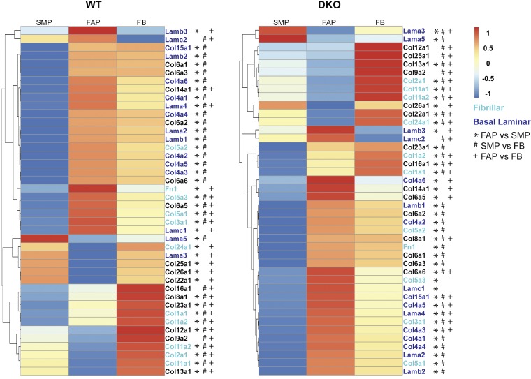
Fig. 8.
Specialization of ECM expression in each of the three cell types during fibrosis. In both WT and DKO mice, collagen I expression is highest in FB followed by FAPs and then SMPs, suggesting that each population does not contribute equally to collagen I expression in both healthy and fibrotic skeletal muscle. Compared with SMP and FAP cells in WT muscle, FBs showed elevated expression of fibrillar ECM proteins, denoted by green lettering. FBs and FAP cells showed elevated expression of basal laminal ECM proteins over SMPs, denoted by blue lettering. Finally, SMP cells appeared to have the lowest amount of ECM expression compared with the other two cell types. Scaled gene expression counts of SMP, FAP and FB cells in DKO animals reveals a distinct role for each cell type in fibrosis. Compared with SMP and FAP cells, FBs showed elevated expression of fibrillar ECM proteins. FAP cells showed elevated expression of basal laminal ECM proteins. Finally, SMP cells had the lowest amount of ECM expression compared with the other two cell types. However, there were a few genes, such as Lama5, that were expressed at a higher level compared with the other cell types. This suggests that these proteins may be important for maintaining the ECM around SMP cells. Symbols depict differentially expressed genes: *differential expression between SMP and FAP, #differential expression between SMP and FB, +differential expression between FAP and FB. Data are presented as row-normalized scaled gene expression counts ranging between −1 and 1.

