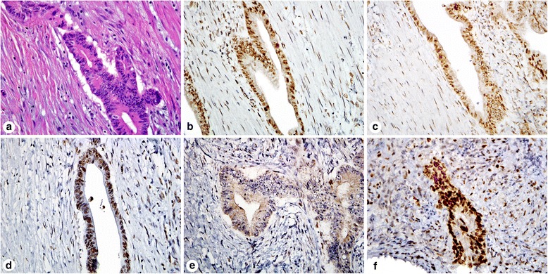
Fig. 4.

Immunohistochemistry for mismatch repair proteins in a patient that received neoadjuvant chemotherapy for rectal adenocarcinoma. H&E stain of the tumor in the resection specimen (a). The resection specimen showed intact MLH1 (b), PMS2 (c), and MSH2 (d) staining. MSH6 staining of the resection specimen showed focal nucleolar staining (e) that was originally interpreted as absent, but subsequent molecular sequencing did not reveal a mutation. The pretreatment tumor biopsy was stained for MSH6 and showed intact staining (f)
