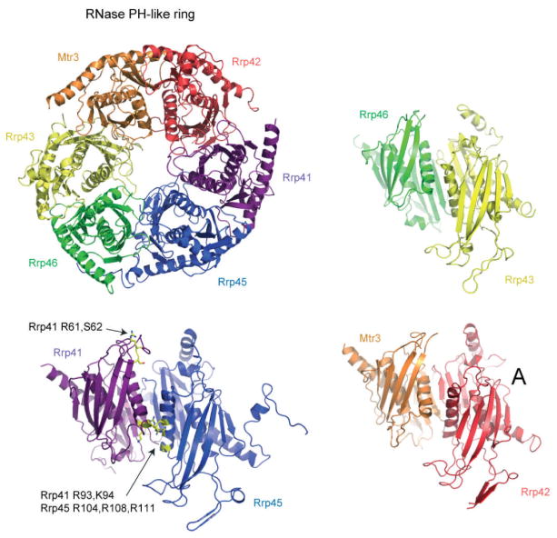
Figure 3.3.
The human PH-like subunit ring. Ribbon diagram of the human PH-like ring (PDB 2NN6) depicting β strands as arrows, α helices as coiled ribbons, and connecting elements as thin tubes (upper left). The view of the intact PH-like ring is from the “top” as presented in Figs. 3.1 and 3.2 with subunits labeled and color coded with Mtr3 (orange), Rrp42 (red), Rrp41 (purple), Rrp45 (blue), Rrp46 (green) and Rrp43 (yellow). The Rrp41/Rrp45 heterodimer is shown lower left in a side view as if the viewer were inside the central channel looking outward. The subunits are labeled and color coded as before with amino acid side chains implicated in RNA-binding interactions labeled and side chains colored yellow for Rrp41 Arg61, Ser62, Arg93, and Lys94 and Rrp45 Arg104, Arg108, and Arg111. The Rrp46/Rrp43 and Mtr3/Rrp42 heterodimers are shown in a similar orientation with subunits labeled and color coded as before.

