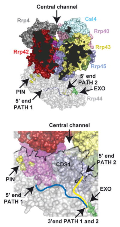
Figure 3.6.

Model of the 10-subunit cytoplasmic exosome. The top panel depicts a side view of the 10-subunit exosome in surface representation that was constructed by aligning Rrp41/Rrp45 from the yeast Rrp41/Rrp45/Rrp44 and human nine-subunit exosome core structures. To enable visualization of the exosome core central channel, the Mtr3 subunit was removed from the complex and parts of Rrp42 and Rrp43 were removed from view (dark areas). The subunits are colored as before except Rrp4 is now depicted in gray. The central channel is labeled and indicated by an arrow at the top of the channel. Rrp44 is depicted in transparent gray with endoribonuclease (yellow) and exoribonuclease (green) sites labeled with side chains in surface representation. Two RNA paths are depicted in Rrp44, one derived from the structure of RNase II in complex with RNA (blue; PDB 2I×1) which passes through the CSDs and S1 domain and the other derived from the dPIN-Rrp44/RNA complex (yellow; PDB 2NVU) which passes by CSD1 and the RNB domain. Bottom panel shows an orthogonal view looking up into the exosome central channel indicating the position of CDS1 which appears to block a direct path from the exosome central channel to the RNB active site (green). RNA paths, PIN and EXO active sites are labeled as in the top panel.
