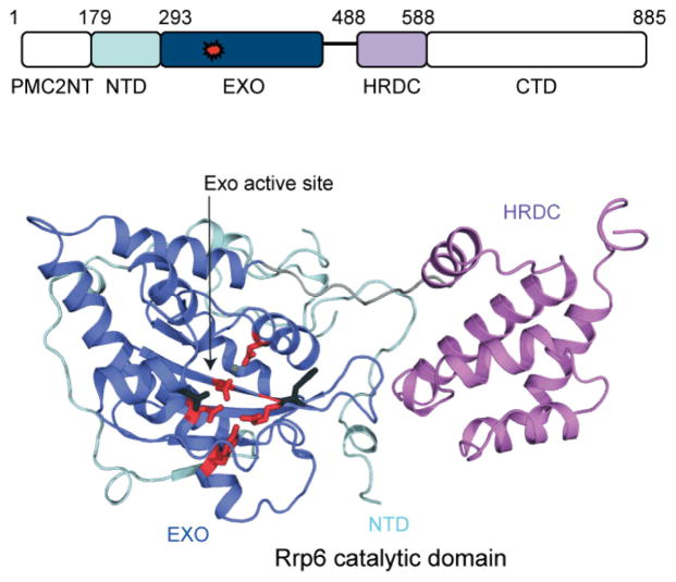
Figure 3.7.
The exoribonuclease Rrp6. Schematic of the Rrp6 polypeptide indicating the PMC2NT (white), NTD (light blue), EXO (dark blue), HRDC (purple) and C-terminal (CTD; white) domains with the exoribonuclease active site colored red. Amino acid numbering is for human Rrp6. Lower panel depicts a cartoon ribbon representation of the human Rrp6 catalytic domain structure with α helices in cartoon ribbon, loops as thin ribbons, and β strands as arrows (PDB 3SAF). Domains are colored and labeled as in the schematic with the linker between the EXO and HRDC domain in gray. The EXO active site is labeled and indicated with an arrow with key side chains colored red and shown in stick representation; the magnesium ion is shown as a small green sphere.

