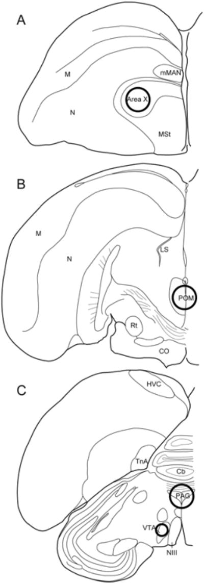
Figure 1.

Coronal sections showing approximate sizes and locations of tissue punches in (A) Area X (2 mm diameter), (B) POM (2 mm diameter), and (C) VTA (1 mm diameter) and PAG (2 mm diameter). Bilateral tissue punches were collected, except for POM and PAG (for each of these regions, a single central punch was taken). Abbreviations: Cb: cerebellum; CO: optic chiasm; HVC: letter-based proper name; LS: lateral septum; M: mesopallium; mMAN: medial magnocellular nucleus; MSt: medial striatum; N: nidopallium; NIII: third cranial nerve; Rt: nucleus rotundus; TnA: nucleus taenia of the amygdala
