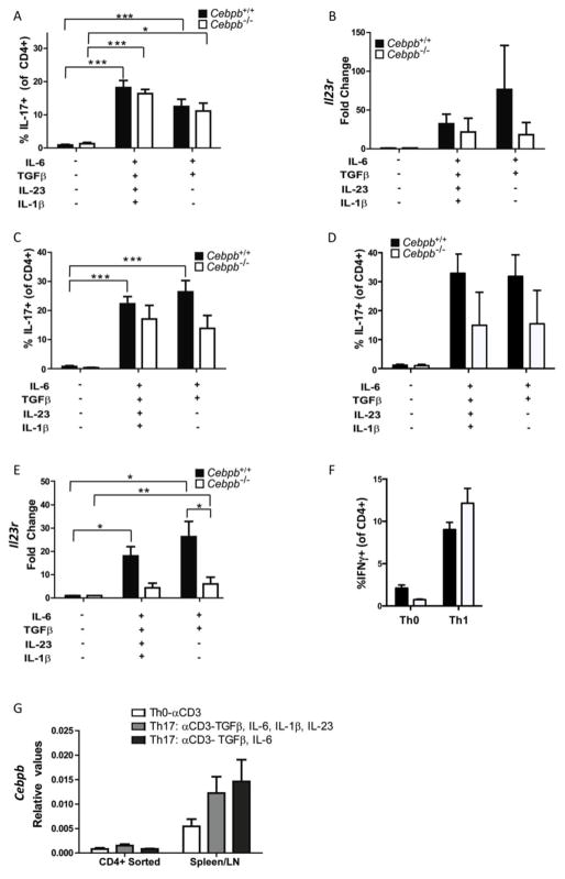
Figure 4. Il23r expression is reduced in Cebpb deficient mice.
A–B: CD4+ T cells were isolated from Cebpb+/+and Cebpb−/− mice and activated with anti-CD3 in presence or absence of Th17-inducing cytokines for 3 days before analysis of IL-17A by flow cytometry (A) and expression of Il23r by qPCR (B). C–D: Total splenocytes/lymph node cells from Cebpb+/+and Cebpb−/− mice were activated with anti-CD3 in presence or absence of Th17-inducing cytokines for 3 days (C) or Th17-inducing cytokines and anti-CD28 (D) before analysis of IL-17A by flow cytometry and expression of Il23r by qPCR (E). F: Total splenocytes/lymph node cells from Cebpb+/+and Cebpb−/− mice were activated with anti-CD3 in presence or absence of IL-12 to induce Th1 cells for 3 days before analysis of IFNγ by flow cytometry. G: Cebpb expression in WT T cells and total splenocyte/lymph node cell cultures. Data are pooled from 2 independent experiments. Bars on graphs show mean and SEM. *P<0.05, **P<0.005 and ***P<0.0005 by ANOVA.

