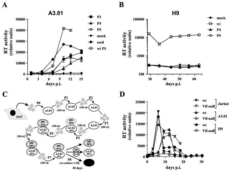
Fig. 5.
Failure to select A3G resistant Vif-null HIV-1 from A3.01 cells.(A) Wild type or NL4-3 Vif-null virus stocks were used to infect A3.01 cells. Virus production was monitored by reverse transcriptase assay. Viruses were passaged 5 times for a combined total of more than 60 days with virus collected at peak virus production serving as input virus for each subsequent passage. Finally, virus from each passage, normalized for reverse transcriptase activity, was used to initiate a parallel infection of A3.01 cells to allow for the identification of changes in viral replication fitness. Vif-null seed virus (HeLa supernatant) and wild type NL4-3 from passage 5 was analyzed in parallel and mock infected cells were cultured alongside (mock). Virus replication was monitored for 15 days and virus production was plotted as a function of time. Analysis of passages P3, P4, and P5 were performed in triplicate and error bars represent the SEM. (B) Vif-null virus from passages 4 & 5 as well as wild type virus from passage 5 were used to infect H9 cells. Virus replication was monitored for 63 days as described for panel A. (C & D) Attempt to isolate A3G-resistant HIV-1 using A3.01/H9 mixed cultures. (C) Schematic outline of the experimental strategy. Virus was initially passaged three times on A3.01 cells (P1-P3) followed by four additional passages on a mixture of A3.01 and H9 cells with increasing ratios of H9 to A3.01 cells (10% to 67% H9 cells). The final passage (P8) shown in panel D was not initiated by cell-free virus but by co-cultivation of mixed cultures from passage 7 (10%) with H9, A3.01 or Jurkat cells (90% each). Overall, the virus was passaged in this experiment for 153 days. (D) Cells from passage 7 were collected 30 days post-infection and co-cultivated with H9, A3.01, or Jurkat cells at a 1:10 ratio. Virus replication was monitored for another 30 days by RT assay and results were plotted as a function of time.

