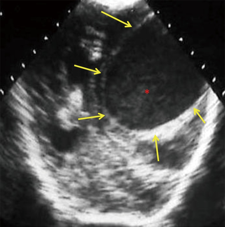
Figure 10.

12-day-old neonate with Citrobacter koseri meningitis. Posterior coronal cranial ultrasound image shows a large well circumscribed hypoechoic cystic lesion (yellow arrows), with echogenic debris/pus (red asterisk), consistent with a brain abscess. Note the mass effect, midline shift, compression of ipsilateral lateral ventricle and dilatation of the contralateral lateral ventricle. The differential includes cystic neoplasm and less likely arachnoid cysts and porencephalic cysts.
