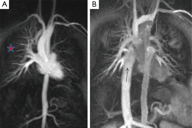
Figure 4.

Cardiac MR angiogram with early (A) and late (B) phase acquisitions. (A) Contrast flows down the right sided SVC to preferentially fill the right pulmonary artery (RPA). There is unilateral opacification of the right pulmonary veins (red star). After contrast has circulated and returns to the heart via the inferior vena cava (IVC) (B), it preferentially fills the left pulmonary artery.
