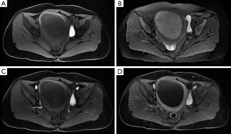
Figure 2.
MRI of the tumor. There is a 7.8 cm × 8.8 cm × 9.9 cm mass tumor in the uterine cavity with distinct margin. The tumor shows slight hypointensity on T1-weighted image (A) and slight hyperintensity on T2-weighted image (B). (C,D) The tumor mass presents slight homogeneous enhancement (C) while the edge of the mass has obvious enhancement (D). MRI, magnetic resonance imaging.

