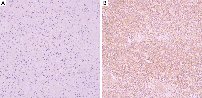
Figure 3.
Pathology and immunohistochemistry. (A) The tumor presents general degeneration and necrosis, nucleus karyopyknosis and sporadic arteriole (×200); (B) immunohistochemistry indicates CD10(+), vimentin(+), ER(+), CD99(±), MPO(−), LCA(−), inhibin(−), PR(−), Syn(−), CD20(−), CD3(−), SMA(−), CgA(−), and ki-67(+, 10%) (×200).

