
Fig. 9.
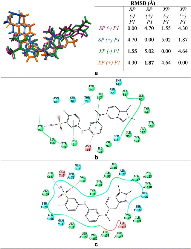
Binding mode of pazopanib in 3VRI. a Alignment of pazopanib from SP and XP scoring functions with and without peptide P1. SP (−) P1 is shown as purple, SP (+) P1 is shown as blue, XP (−) P1 is shown as green, and XP (+) P1 is shown as orange. b 2D binding mode from SP (−) P1. c 2D binding mode from SP (+) P1