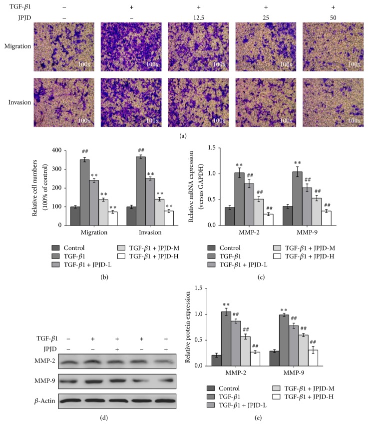
Figure 3.
Impact of JPJD on the invasion and metastasis of TGF-β-stimulated LoVo cells. (a) Transwell experiment was performed to observe the impact of JPJD on the invasion and metastasis of TGF-β-stimulated LoVo cells. The magnification of the microscopic pictures was ×100. (b) Numbers of invasive and migrated cells shown as mean ± SD, n = 3. (c, d, e) Real-time PCR, western blot, and ELISA were, respectively, performed to test the effect of JPJD on the MMP-2 and MMP-9 expression of TGF-β-stimulated LoVo cells, as well as the secretory levels of MMP-2/MMP-9 in the culture medium of TGF-β-stimulated LoVo cells. Here, JPJD-L is 12.5 μg/mL, JPJD-M is 25 μg/mL, JPJD-H is 50 μg/mL, and the time of TGF-β-stimulation and JPJD treatment was 48 hours. ##P < 0.01, versus control LoVo cells, and ∗P < 0.05 and ∗∗P < 0.01, versus only TGF-β-stimulated cells.

