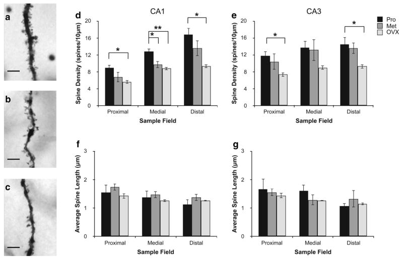
Fig. 6.
Effects of stage of the estrous cycle and ovariectomy on dendritic spines in the CA1 and CA3 regions of the rat hippocampus. Brightfield micrographs (×630) of sections of apical CA3 dendrites in the stratum radiatum from proestrous (Pro) (a), metestrous (Met) (b) and ovariectomized (OVX) (c) females. Scale bars 10 μm. Spine density (d) and length (f) in the proximal, medial and distal dendrite segments (see “Methods and materials” for details) of CA1 pyramidal neurons, from Pro, Met, and OVX rats. e, g Corresponding data from CA3 neurons. Statistical analysis: a significant overall treatment effect [ANOVA (reproductive state × dendritic field); F(2,42) = 23.001, p < 0.0001] and interaction effect between dendritic field and hippocampal region (ANOVA; F(2,42) = 3.502, p = 0.0405) was observed. OVX females had significantly reduced dendritic spine density compared to Pro females across all fields examined, with the exception of the medial dendrite segment of CA3. Significant differences between proestrus and metestrus animals were also observed in the medial segment (corresponding approximately to stratum radiatum) in CA1, but not CA3. Dendritic spine length did not differ significantly between the three groups (ANOVA; F(2,42) = 1.038, p = 0.3629). Data represents mean ± SEM (n = 3–4 per group)

