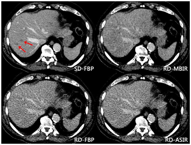
Figure 3. False negative lesions at reduced dose reconstructions.
Red arrows indicate metastatic lesions within the right lobe of the liver on standard dose FBP imaging [top left] of an 82-year-old male with metastatic neuroendocrine tumor. These lesions were not identified at reduced dose MBIR [top right], FBP [bottom left] or ASIR [bottom right]. Dose reduction was 74% on the reduced dose series, with an effective dose of 1.49 mSv. Additional lesions in the left lobe are partially seen on the standard dose image but not clearly seen on the reduced dose series.

