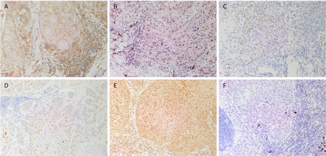
Figure 1.
Representative DAB immunohistochemical-stained sections of moderately differentiated lip squamous cell carcinoma demonstrating nuclear membrane staining of CD44 [(A), brown] on cells within the stroma and cell membrane staining on cells within the tumor nests (TNs). Nuclear staining of NANOG [(B), red] was seen in cells within the TNs and the stroma. Patchy areas of weak staining for phosphorylated signal transducer and activator of transcription 3 [(C), brown] was detected on cells within the TNs. Focal moderate expression of spalt-like transcription factor 4 [(D), brown] on cells within the TNs and weak staining on cells within the stroma. Widespread and strong staining of sex-determining region Y-box 2 [(E), brown] was seen on cells within the TNs and the stroma. Staining for octamer-binding transcription factor 4 [(F), red] was limited to cells within the stroma. Original magnification: 200×.

