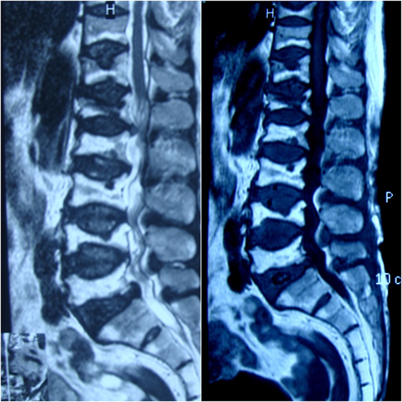
Figure 2.

Sagittal T1-weighted (right) and T2-weighted (left) MRI of the lumbosacral spine. The D10–12 vertebrae are also seen. All vertebral bodies show biconcave deformity of their endplates with areas of rounded depressions and impressions, different signal intensities of the vertebral body and bone marrow, and loss of more than 40% of the vertebral height. This is severe osteoporosis showing different stages of bony collapse.
