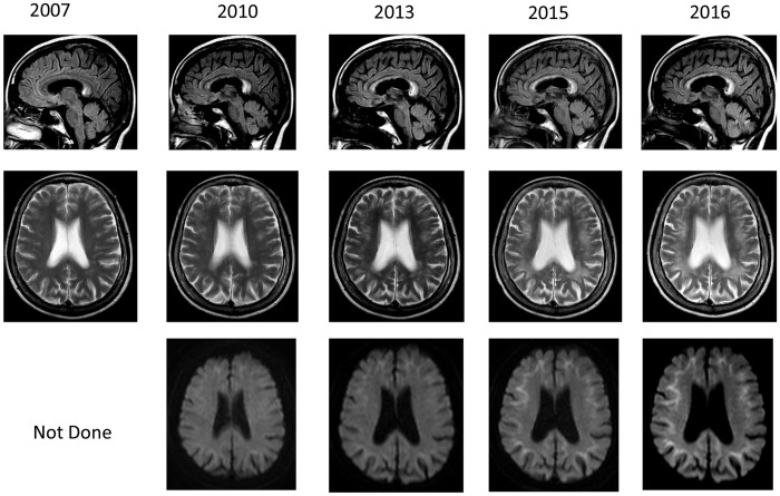
Figure 1.
Brain MRI findings. Brain MRI findings showed leucoencephalopathy in T2-weighted and fluid attenuation recovery (FLAIR) images, and showed high-intensity lesions in the corpus callosum. On diffusion-weighted images (DWIs), high intensities were observed from the corticomedullary junction to around the root of gyrus. These abnormal MRI findings were gradually expanded from the frontal to the occipital that were more prominent in DWIs.

