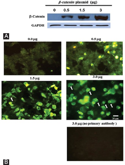
Figure 1.

HEK293T cells were seeded in duplicate and transfected with different amounts of the β-catenin plasmid (the number on top of each panel). One group of cells was used for western blotting experiments to measure β-catenin protein levels (A) and the other group was used for immunofluorescence staining of β-catenin (B). The cells harboring β-catenin protein aggregates are indicated by arrows. The lowest panel represents the cells transfected with 3 μg of the β-catenin plasmid, but the primary antibody was omitted from the staining protocol to test the specificity of the β-catenin antibody. The expression of the GAPDH protein was used as a loading control for the blot shown in figure 1A.
