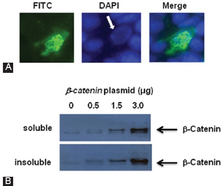
Figure 2.

(A) FITC (left) and DAPI (middle) staining of a HEK293T cell, overexpressing β-catenin. The figure shows that the β-catenin protein aggregates predominantly formed in the cell nucleus (the arrow). The right hand panel is the merged image. (B) HEK293T cells were transfected with increasing amounts of the β-catenin plasmid; and 48hours after transfection, the cells were harvested and fractionated as described in Reference 16. Nuclear soluble and insoluble proteins were utilized for immunoblotting experiments using β-catenin antibody.
